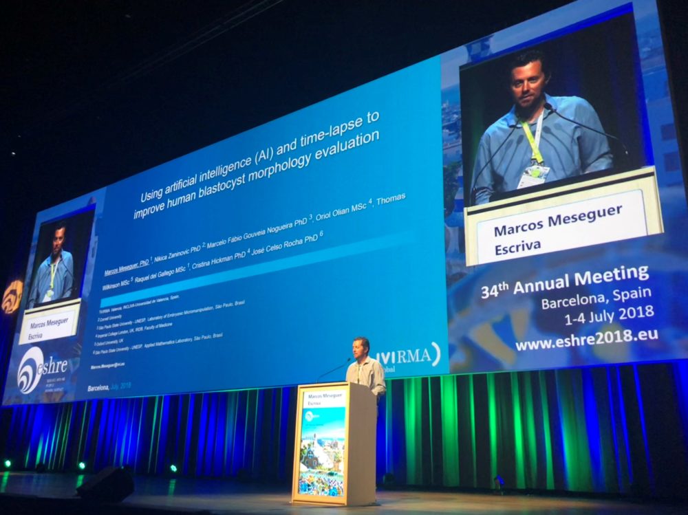
As part of the 34th Congress of the European Society of Human Reproduction and Embryology (ESHRE):
- This technology is much faster and more consistent than an embryologist in classifying embryos using Time-Lapse imaging
- Its use can significantly improve our ability to assess embryo viability
BARCELONA, JULY 4, 2018.
Historically, the role of the embryologist has been to assess embryos’ morphology and growth rate and select the one(s) most likely to result in a pregnancy for transfer into the maternal uterus in women treated with assisted reproductive technologies. This reality is the key starting point of the study entitled “Using Artificial Intelligence (AI) and Time-Lapse to improve human blastocyst morphology evaluation”, presented by Dr. Marcos Meseguer, an embryologist at IVI Valencia, at the 34th Congress of the European Society of Human Reproduction and Embryology (ESHRE), in Barcelona.
“This research shows how Artificial Intelligence (AI) is much faster and more consistent than the embryologist in classifying embryos using time-lapse imaging versus the variability and heterogeneity linked to the work of the human operator alone”, explains Dr. Meseguer, lead author of the study.
This is how he summarizes the conclusions of the study, where the Universidade Estadual Paulista (UNESP) participated, and 5 embryologists from 4 different countries analyzed 223 embryos using conventional morphologic criteria for embryo selection. AI then was used to learn to measure, interpret, analyze, and distinguish the different aspects of embryo development and to select viable embryo(s) according to these criteria, perfecting its process as the number of assessed embryos increased.
“The application of AI to the classification of the human blastocyst is economical, non-invasive and more reliable than the classification by a human operator. Instead of a human looking at thousands of images, the AI continuously evaluates, quantifies, learns and re-evaluates. As demonstrated, this technology can improve our ability to assess embryonic viability,” he added.
To achieve higher precision in the evaluation of embryo viability and to reduce the subjectivity that affects the process of embryo selection, digital image processing and AI techniques were used in conjunction with Time-Lapse imaging, allowing Dr. Meseguer and his colleagues to accurately determine the appropriate time-points for embryo assessment; introducing consistency into the process. This analysis was carried out identically in different sites since it was performed based on fixed timing and standardized Time-Lapse images. However, it is noteworthy that human blastocysts present challenges for image analysis using AI and further independent studies are required to demonstrate reproducibility of the reported finding prior to wider clinical application.


