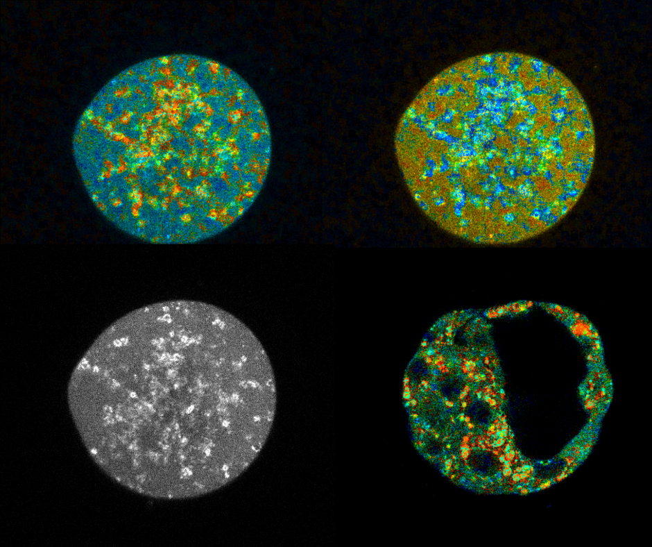
- FLIM (fluorescence-lifetime imaging microscopy) is a live microscopy technique based on the use of fluorescence in real time
- Dr. Emre Seli, Research Director of the IVIRMA Group, has conducted a study in which, oocyte metabolic function was assessed using FLIM
- This new technology, which reveals metabolic data from mitochondria, is providing promising results as the alternative to current invasive techniques of mitochondrial function assessment
VALENCIA, JANUARY 7 th, 2019.
Oocyte development, fertilization, and pre-implantation embryo development have unique energy requirements that depend on timely changes in mitochondrial activity and structure. Accordingly, through the past few decades, mitochondria have been investigated as diagnostic and therapeutic tools in assisted reproduction.
In a study lead by Dr. Emre Seli, the Research Director of the IVIRMA Global, in collaboration with Drs. Dan Needleman and Tim Sanchez from Harvard University, a new microscopic approach called FLIM (fluorescence lifetime imaging microscopy) was shown to effectively assess the metabolic state of oocytes.
According to Dr. Seli, “This is a very exciting technology that is non-invasive and provides information in real time. For the time being, it is only applied for research purposes. However, I can’t help being excited about its potential applications in clinical IVF”.
NADH and FAD play central roles in cellular energy production by carrying electrons to the electron transport chain. Importantly, NADH and FAD are fluorescent molecules, which means that shining light on them of one wavelength can cause them to transition to an excited state and emit light of another wavelength as they relax back to their original state. By measuring the amount, locations, and protein binding status of these molecules, FLIM allows real time analysis of the metabolic state of an oocyte or embryo.
In the current study (Metabolic Imaging with the use of fluorescence lifetime imaging microscopy (FLIM) accurately detects mitochondrial dysfunction in mouse oocytes), Dr. Seli and his colleagues used FLIM to compare the metabolic state of oocytes with mitochondrial dysfunction (as a result of targeted gene deletion) to normal oocytes. There was a very significant difference in the parameters detected by FLIM. Similarly, they compared oocytes from older mice to those from young mice; indeed metabolic differences were significant. Importantly, changes in mtDNA copy number, a mitochondrial parameter recently applied clinically, was less prominent and not significant in comparing old vs young.
Therefore, although Dr. Seli emphasizes that this new technique is not yet ready for clinical application, it is already emerging as a promising tool for mitochondrial assessment, especially for research studies.


