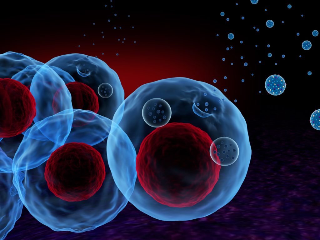
Communication between cells is paramount for adequate cell function. Cells in our body are constantly sending and receiving messages that are translated into various cellular responses and sometimes complex biological processes. These messages consist of molecules that are secreted by cells in very different ways. One of the most efficient and well documented mechanisms of cellular communication is extracellular vesicles. Extracellular vesicles (EVs) are membraned structures secreted by cells that contain molecules and potential messages in their lumen and that can carry them through long distances until getting in contact with other recipient tissues. The term extracellular vesicle refers to a highly heterogeneous population of cellular couriers that differ vastly in size and function. Exosomes, microvesicles and apoptotic bodies are some examples of the terminology used to describe different subpopulations of EVs.
Given the pivotal role played by EVs in cellular communication and cell function, it is highly likely that these vesicles are important key regulators of human reproduction. In fact, it has been thought that by studying EVs, their nature and the messages they convey, we could shed light on several reproductive processes that we still do not fully understand, not to mention the potential of improving clinical outcomes.
Several studies have given solid proof of the existence of these EVs is various body fluids critical for reproduction such as follicular fluid, endometrial fluid, semen and even fluid in the Fallopian tubes [1, 2]. For instance, major populations of EVs have been described in seminal plasma [3]. Although their function still remains inconclusive, some recent reports found that exposure of isolated seminal vesicles to poor quality sperm could boost their reproductive potential, suggesting a promising field of research on sperm EVs [4]. Furthermore, other studies have found that molecules carried by EVs in follicular fluid are associated with IVF outcomes and could be used as predictors of a cycle’s success. However, our group has payed special attention to the molecular dialog between the embryo and endometrium, since this process is indispensable for correct embryo implantation and pregnancy.
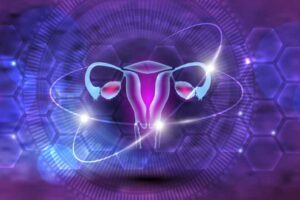
Embryo-endometrium crosstalk
When an embryo is in the uterus, either by natural conception or after an embryo transfer, it engages in a series of events that hopefully lead to embryo implantation and the beginning of a pregnancy. During those first moments of contact with the endometrium, both the maternal tissue and the embryos outer cells secrete and receive cues and stimuli that prepare both parts for the process of implantation. Endometrial EVs have been very well described both in structure and content. Embryo-derived EVs, on the other hand, are much more challenging to study given that only the embryo itself produces them, in contrast to an entire endometrial lining with millions of cells.
Therefore, our IVF labs are the perfect setting to investigate embryonic EVs, since we keep our embryos in microdrops of culture medium until they become blastocysts. That culture medium can be later analysed for the presence of these entities. Recently and after testing several methodologies, our group has successfully isolated EVs from these spent culture medium microdrops, making the first step to understand how important these vesicles are for embryo biology and the process of implantation. In addition, our data suggest that EVs are already present in the culture medium unexposed to the embryos! Such a finding can even change the way at how we look at culture media, and can open the door for a myriad of new exciting research questions about EVs of non-embryonic origin [5].
Although there are still many technological challenges to overcome in order to optimize embryonic EV isolation, it now seems more possible than ever to start to unravel the secrets that these vesicles might be keeping. For example, should it be discovered that some embryos differ in their rate of production of EVs, or in the molecules that EV carry, EVs could become indicators of embryo biology and viability, setting the grounds for another way to improve embryo selection in an IVF cycle.
EVs have also proved to be of great importance in the field of cancer biology, since these small “packages” can be engineered to carry and deliver their cargo to specific cells. In addition, they are extremely helpful in diagnosing diseases at early stages. These discoveries together with the available material we have in the IVF lab encourage us to further explore the potential that EVs have to help us understand some of the black boxes in reproductive medicine, as well as their use as biomarkers of embryonic competence.
- Simon C, Greening DW, Bolumar D, Balaguer N, Salamonsen LA, Vilella F. Extracellular Vesicles in Human Reproduction in Health and Disease. Endocrine reviews. 2018;39(3):292-332.
- Machtinger R, Laurent LC, Baccarelli AA. Extracellular vesicles: roles in gamete maturation, fertilization and embryo implantation. Hum Reprod Update. 2016;22(2):182-93.
- Hoog JL, Lotvall J. Diversity of extracellular vesicles in human ejaculates revealed by cryo-electron microscopy. Journal of extracellular vesicles. 2015;4:28680.
- Murdica V, Giacomini E, Alteri A, Bartolacci A, Cermisoni GC, Zarovni N, et al. Seminal plasma of men with severe asthenozoospermia contain exosomes that affect spermatozoa motility and capacitation. Fertil Steril. 2019;111(5):897-908.e2.
- Marin D, Seli E, Scott R. Extracellular Vesicles Can Be Isolated From Culture Media With And Without Exposure To Human Preimplantation Embryos. 8th International IVIRMA Congress 2019; April, 2019; Palma de Mallorca, Spain.2019.
Author: Diego Marin